
Respiratory System Anatomy – Major Zones & Divisions
The respiratory is separated into the conducting zone and the respiratory zone.
The conducting zone include structures that facilit air to move in and out of the lungs.呼吸器は、導電部と呼吸部に分かれています。
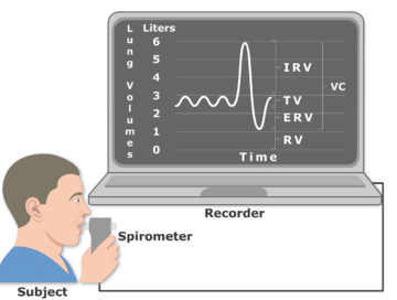
Lung Volumes and Capacities
肺内の空気量は、4種類の容量と4種類の容積に細分化されることが可能です。 4つの容量とは、潮容積、吸気予備量、呼気予備量、残量である。 4つの容量には、吸気容量、機能的予備容量、生命維持能力、全肺容量があります。
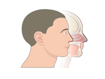
鼻と鼻腔|構造 & 機能
鼻と鼻腔についてご紹介しています。 鼻は呼吸器の最初の部分であり、空気交換のための通気口としての役割を担っている。
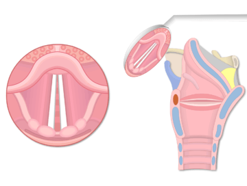
Glottis – Structure & Function
声帯とその間の空間を声帯と呼びます。 喉頭筋は、声門の開口部の大きさを調節します。 声帯の開口部が広ければ、気管への空気の出入りが容易になります。
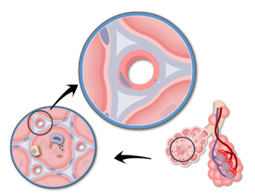
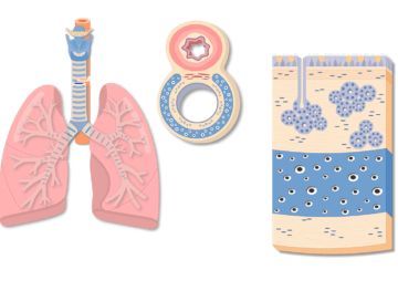
Respiratory Membrane and Gas Exchange
呼吸膜の構造および肺で起こるガス交換のメカニズムに関するインタラクティブなデモを行います。
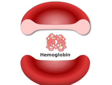
Hemoglobin Molecule – Structure & Function
Hemoglobin の構造を詳細かつインタラクティブに、その状態(リラックス状態 & 緊張状態)についてカラフルでシンプルなアニメーションを使用して説明します(グロービン、アルファおよびベータ サブユニット、ポルフィリン、ヒームグループ、…など)。
気管壁の構成と構造-気管チューブまたは気管の解剖学
気管壁の組織は4層になっています。 気管には呼吸粘膜、粘膜下層、軟骨輪、気管筋、外膜があります。
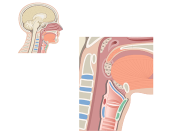
Larynx の位置と機能
喉頭は咽頭と気管の間の空気の通路で保護されている。 9つの支持軟骨、固有筋と外筋、粘膜の裏打ちで形成されている。 喉の中で舌骨と舌の下、食道の前方に位置する1.5インチの短い管である。
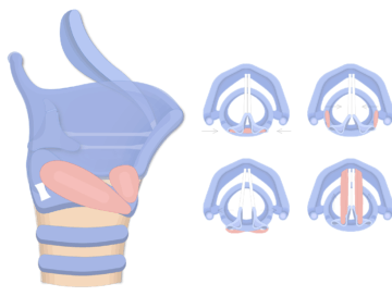
喉頭固有筋
固有筋は、アリテノイド軟骨を動かし、声帯や靭帯にかかる張力を調節する働きがあります。
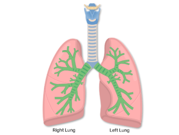
気管支の構造、機能、&位置|Bronchus Anatomy
胸骨角付近で気管が分岐して、左右一番気管支(①)に分かれている。 それぞれの気管支は数cmの距離を自由に走行した後、それぞれの肺に入る。 空気は、この気管支を通じて、それぞれの肺に出入りしている
。